Weill Cornell Medicine employs the latest scientific advances in cancer care, from precision medicine and immunotherapy, to minimally invasive procedures and robotic surgery.
Each week, our cancer specialists gather for a Thoracic Tumor Board, where they discuss their cases and collaborate on the best ways to treat each patient. The tumor board includes thoracic surgeons, medical oncologists, radiation oncologists, pulmonologists, pathologists, interventional pulmonologists and interventional radiologists.
While individual care plans vary — based on a person’s specific needs and circumstances — patients typically receive a combination of the following treatments and services.
Lung Cancer Screening Program
Health experts recommend lung cancer screenings for high-risk individuals. At the Lung Cancer Screening Program at Weill Cornell Medicine and NewYork-Presbyterian, patients receive comprehensive treatment from a team of experts, from diagnosis to treatment, if necessary.
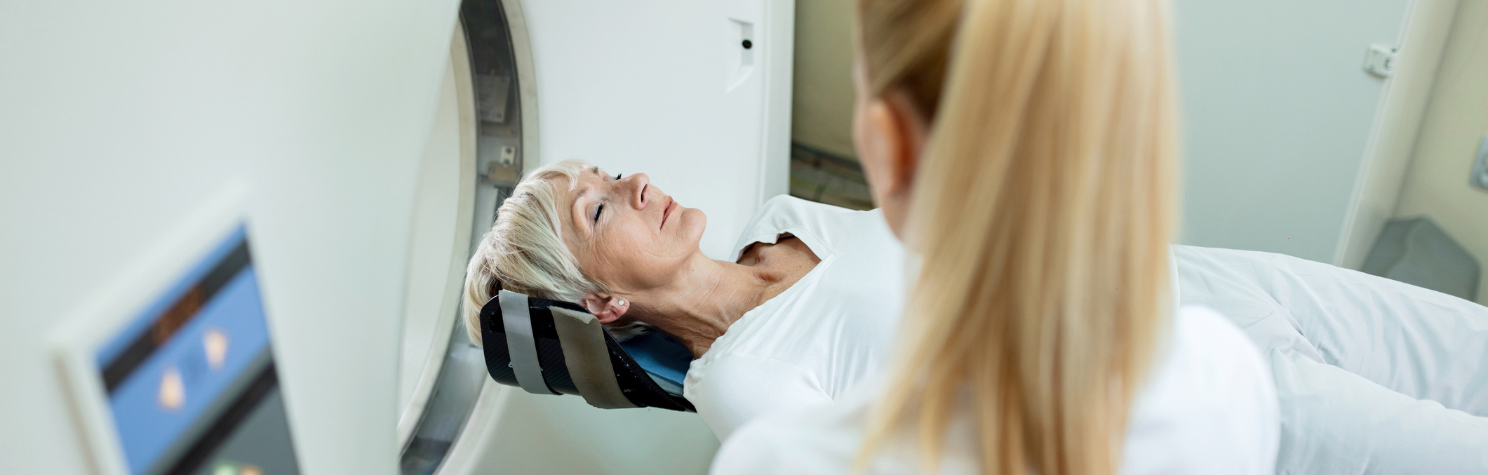
In alignment with the U.S. Preventative Services Task Force (USPSTF) recommendations, the Lung Cancer Screening Program provides annual screening using low-dose computed tomography (CT) to check for signs of lung cancer in high-risk individuals.
Individuals who meet all the following conditions are considered at “high-risk” by the task force:
55 to 80 years old
Current or former smokers who have quit within the last 15 years
Have a history of heavy smoking, equivalent to at least 30 pack years
No signs or symptoms of lung cancer
A pack-year equals the number of packs smoked per day times the number of years the patient smoked. For example, 1 pack per day for 30 years equals 30 pack-years.
Those who do not fit these guidelines, but who may have other factors that put them at risk for lung cancer — such as an immediate family member who was diagnosed with lung cancer at an early age — can also receive personalized assessments. While individuals who do not meet these guidelines may not receive insurance coverage for the exam, these assessments can help them decide if they want to undergo a low-dose screening CT.
Following their scan, patients have the option to review their images immediately after with a radiologist. In addition to screenings, the program can help patients coordinate follow-up care. The Lung Cancer Screening program also offers a range of support groups for participants and their families, including tobacco treatment counseling.
Chemotherapy, Precision Medicine and Immunotherapy
Weill Cornell Medicine provides compassionate care informed by a deep expertise in lung and other cancers of the chest.
The oncologists in the Thoracic Medical Oncology Program have dedicated their careers to treating these diseases exclusively, while conducting research to develop new and better treatments. They are highly regarded in the field, with expertise in:
Targeted therapies
Immunotherapy
Tumor genetics
Treating advanced (Stage 3 and Stage 4) lung cancers
Weill Cornell Medicine also offers a number of clinical trials, giving patients access to new and advanced treatments that may not be available anywhere else.
Our doctors also prioritize their relationships with patients, taking the time to get to know them. They partner with each person they treat to determine the best evaluation and treatment program for them, whether that be through standard of care therapies or participation in one of our clinical trials.
Chemotherapy
Chemotherapy drugs kill cancer cells directly. While some people may only receive chemotherapy to treat their cancer, most use it in combination with other treatments.
Because each person we treat receives a personalized care plan, the combination may vary, but chemotherapy can be used to:
Make a tumor smaller before surgery (neoadjuvant chemotherapy)
Destroy cancer cells that may remain after surgery or radiation therapy (adjuvant chemotherapy)
Help other treatments such as radiation or immunotherapy work better
Kill cancer cells that have returned or spread to other parts of the body.
At Weill Cornell Medicine, patients receive chemotherapy in comfortable and modern infusion centers. Our infusion centers are conveniently located in the same building as our doctors’ offices, making it easier to receive comprehensive care.
Precision Medicine and Targeted Therapies
Weill Cornell Medicine is at the forefront of Precision Medicine, a relatively new approach in the fight against cancer.
Unlike traditional chemotherapy – which kills all rapidly growing and dividing cells – Precision Medicine targets a specific pathway or process in a tumor. The goal is to impair the tumor’s ability to grow, divide and spread. Since Precision Medicine targets cancer cells specifically, normal cells are less affected, thereby reducing side effects like hair loss and low blood cell counts.
Precision Medicine is a highly personalized approach to treatment. Our team takes a sample of the patient’s tumor and analyzes it to identify specific targets for treatment. Unlike many other institutions, we conduct this analysis in-house, allowing our oncologists to communicate directly with the molecular pathologists running the tests who can provide more insights about the results.
Immunotherapy uses the body’s natural defenses to fight cancer. The immune system helps your body fight infections and other diseases. However, it often has a tough time targeting cancer cells, which avoid detection by finding ways to alter the immune environment.
Some immunotherapies work by helping the immune system recognize cancer cells. Others strengthen the immune system’s response to cancer cells so it can destroy them.
Weill Cornell Medicine is a leader in immunotherapy, with a robust research program. In addition to standard treatment options, we offer clinical trials that evaluate new immunotherapies and innovative ways of combining immunotherapy with other cancer treatments.
Weill Cornell Medicine is home to one of the most respected thoracic surgery programs in the world.
Our thoracic surgery program is also one of the busiest programs globally, which speaks to our expertise in the field. Unlike institutions with lower patient volumes, our surgeons have treated a wide range of conditions — from straightforward cases to complex surgical problems of the lung, esophagus and thymus.
This expertise, combined with our personalized approach to care, leads to surgical outcomes that are consistently outstanding.
Minimally Invasive Surgery at Weill Cornell Medicine
The majority of our lung and thoracic cancer patients, about 90%, undergo surgeries that use a minimally invasive approach.
Unlike traditional “open surgery” — which includes a large incision in the chest and abdomen — minimally invasive techniques use small, more discreet incisions. As a result, patients experience less pain and faster recovery.
At Weill Cornell Medicine, our minimally invasive procedures include video-assisted thoracoscopic surgery (VATS) and robotic surgery.
Video-Assisted Thoracoscopic Surgery (VATS)
In this procedure, surgeons make small incisions in the chest and/or abdomen. Video-cameras are placed through these tiny incisions so surgeons can visualize the organ on a screen. Small instruments manipulate and remove the tumor and surrounding tissues.
After thoracoscopic surgery, most patients need to stay in the hospital for only 1-3 days. Patients who have thoracoscopic surgery also usually require fewer pain medications than those who have traditional, “open” surgery and enjoy a faster return to normal daily activities.
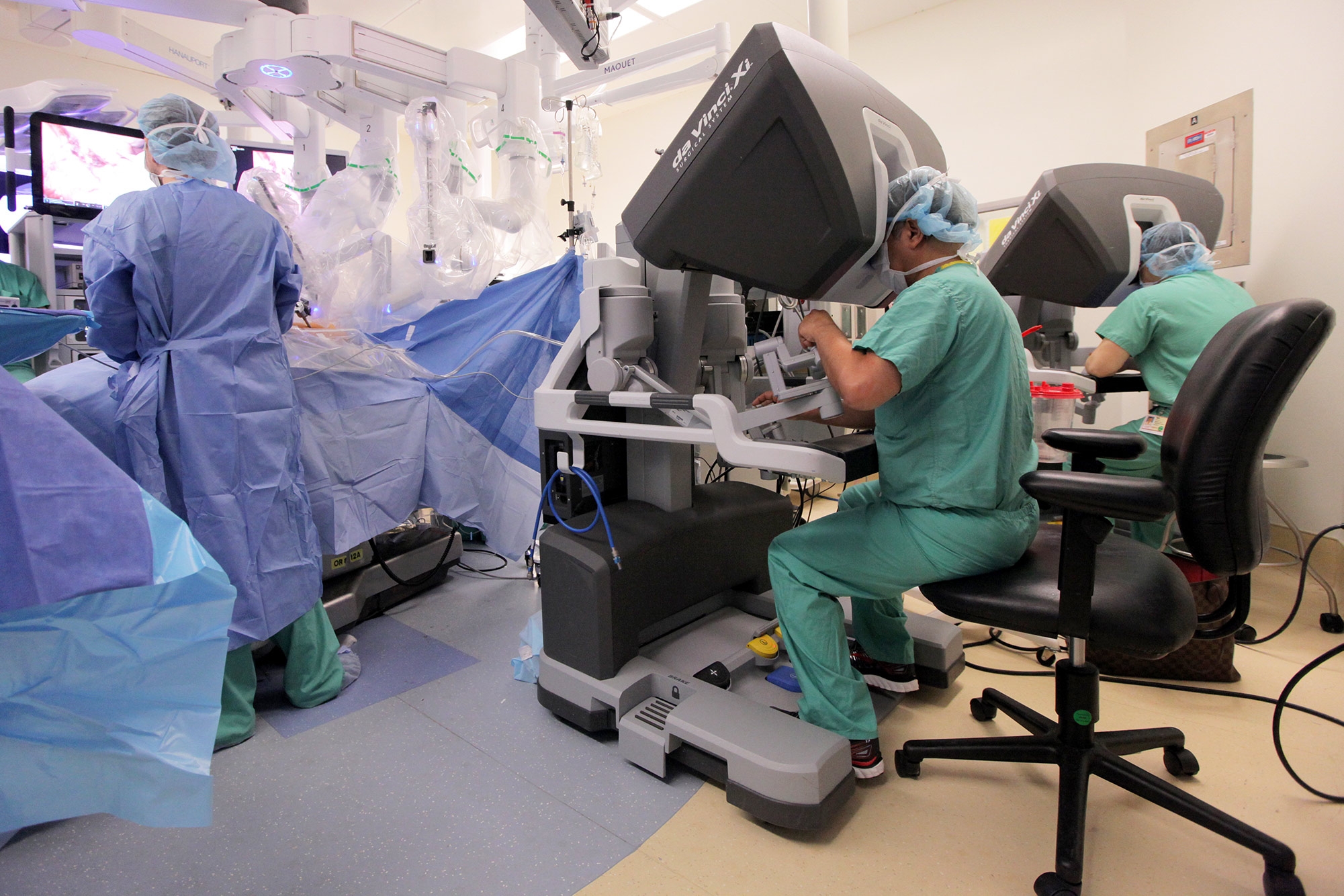
Robotic Surgery
In some cases, surgeons can remove tumors using a robotic approach. During the procedure, the surgeon sits at a console, viewing the surgical field through the robot's "video system."
The surgeon uses controls to operate the various arms and cameras of the robot, which are placed into position in the patient by surgical assistants. The robot enables the surgeon to operate with greater magnification of the surgical field and with more precision in the use of surgical instruments.
Lung-Sparing Surgery
In addition to these minimally-invasive procedures, Weill Cornell Medicine offers lung-sparing surgery. In this procedure, surgeons remove the tumor while preserving as much of the lung as possible.
This approach is recommended for adults with Stage I non-small cell lung cancer and who are more than 75 years old or who have other health conditions that may make lobectomy more complex.
Interventional Pulmonology
Interventional Pulmonology uses the latest technology to diagnose and treat a variety of lung conditions using minimally invasive techniques. As a relatively new subspecialty within pulmonary medicine, interventional pulmonology is often at the forefront of developing and using new technology.
Interventional pulmonologists help lung and thoracic cancer patients in a variety of ways and can be an important part of a patient’s care team. Instances where interventional pulmonologists maybe involved include:
Major airway blockages caused by tumors
Pleural-space diseases, which are a common problem when cancer has spread to the chest
Biopsy of small lung nodules in early stage lung cancer
Mediastinal staging of non-small cell lung cancer, which helps doctors determine how far the disease has progressed
Management of treatment-related side effects, such as post-radiation or post-immunotherapy pulmonary complications
Using advanced endoscopic techniques, Weill Cornell Medicine’s Interventional Pulmonologists perform procedures that were traditionally not possible or required extensive surgery. They provide personalized, expert care through the Advanced Bronchoscopy Program and the Complex Airway Disease Program, which both use cutting-edge tools and techniques.
Radiation therapy uses high-energy X-rays to destroy cancer cells and shrink tumors. Radiation can be used alone or in combination with other treatment options, such as surgery or chemotherapy.
Radiation therapy is also used to provide relief when a cancer has been deemed incurable. By shrinking a tumor, radiation therapy can improve a patient’s quality of life by reducing pressure, bleeding, pain or other symptoms.
Weill Cornell Medicine is a leader in the field, with extensive expertise in radiation therapy.
We also provide personalized care to each person we treat. Our radiation oncologists regularly check in with their patients to follow their progress, to evaluate and recommend treatments for side effects and to address any concern they may have.
We offer both external beam radiation therapy and brachytherapy for the treatment of lung and thoracic cancers.
External Beam Radiation Therapy
External beam radiation therapy destroys tumors and nearby cancer cells by using beams of high-energy photons, electrons or protons. Typically, patients sit or lie down on a couch while an external source of radiation pointed at a particular part of the body.
Radiation oncologists at Weill Cornell Medicine and NewYork-Presbyterian also have access to an MRI guided linear accelerator, which was the first installed in the New York area. With this device, tumor motion is visualized as treatment is being delivered, making treatment even more accurate.
At Weill Cornell Medicine, we offer several types of external beam radiation therapy, including:
Image-Guided Radiation Therapy (IGRT)
Image-guided radiation therapy uses imaging during radiation treatment to improve the precision and accuracy of the treatment delivery.
Using a special imaging system for IGRT, our team creates images of the tumor and the surrounding area. We compare those new images with those taken during the simulation scans and adjust, as necessary, to more precisely target the tumor and avoid surrounding healthy tissue.
Intensity-Modulated Radiation Therapy (IMRT)
Intensity-modulated radiation therapy (IMRT) uses multiple small radiation beams to deliver precise doses of radiation to cancerous tumors.
Using computer-controlled linear accelerators, IMRT controls the intensity of the radiation beam in small volumes. By doing so, the dose can conform more precisely to the three-dimensional share of the tumor.
The procedure’s precise nature also allows physicians to more accurately target the tumor while sparing as much of the surrounding healthy tissue as possible. As a result, the possibility of radiation-induced side effects is significantly lower, compared to non-modulated, or controlled, 3D-conformal radiotherapy techniques.
Due to its complexity, IMRT needs slightly longer treatment times than conventional radiotherapy. Longer planning time and additional safety checks are also required before treatment starts to make sure all planning data are properly transferred from the treatment planning system to the treatment machine and to ensure all modulated beams can be accurately delivered.
Volumetric Modulated Arc Therapy (VMAT)
Volumetric modulated arc therapy (VMAT) is an advanced form of intensity-modulated radiation therapy (IMRT).
VMAT delivers radiation beams by equipment that continuously rotates around the patient, drastically shortening treatment time compared to more conventional radiation treatment. Each rotation is called an arc and one or more arcs might be used.
During each rotation, the radiation is delivered to the intended area only, allowing the radiation oncologist to spare more surrounding healthy tissue.
Both VAMT and IMRT are equally effective for normal tissue sparing. However, the treatment time for VMAT is significantly shorter and thus benefits patients who require longer (30 minutes or more) treatment time.
Stereotactic Body Radiation Therapy (SBRT)
Stereotactic Body Radiation Therapy (SBRT) uses precisely-targeted radiation to treat tumors. It delivers focused radiation in higher doses than typical radiation therapy, but in fewer treatments, thereby shortening the treatment course.
SBRT can be a convenient option for some patients. Its non-invasive nature also makes it a good alternative for patients who are unable to undergo surgery, or who have tumors that are hard to reach or located near vital organs.
At Weill Cornell Medicine, our radiation oncologists plan extensively to ensure that the pencil-thin radiation beams destroy tumors and cancer cells, while avoiding normal tissue.
Brachytherapy
Brachytherapy, also called internal radiation or seed implants, is the placement of radioactive sources in or just next to a tumor. The radioactive sources may be left in place permanently or only temporarily, depending upon your cancer. To position the sources accurately, special catheters or applicators are used.
The powerful radioactive sources travel to the tumor through small tubes called catheters. They travel for the amount of time prescribed by a radiation oncologist.
Radiation oncologists can complete brachytherapy in about 10 to 20 minutes. You may be able to go home shortly after the procedure. Depending on the area treated, you may receive several treatments over a number of days or weeks.
There are two main types of brachytherapy: intracavity treatment and interstitial treatment.
Intracavity Treatment – With intracavity treatment, the radioactive sources are put into a space near where the tumor is located, such as the cervix, the vagina or the windpipe.
Interstitial Treatment – With interstitial treatment, the radioactive sources are put directly into the tissues, such as the prostate.
Often, these procedures require anesthesia and brief hospitalization. Patients with permanent implants may have a few restrictions at first and then can quickly return to their normal activities.
However, most patients feel little discomfort during brachytherapy. If the radioactive source is held in place with an applicator, you may feel discomfort from the applicator. There are medications that can help this. If you feel weak or queasy from the anesthesia, your radiation oncologist can give you medication to make you feel better.
Interventional Radiology
Interventional radiology procedures may be good options for treating and managing lung or thoracic cancers, especially for those who are not able or do not want to undergo surgery.
At Weill Cornell Medicine, our interventional radiologists are highly qualified and experienced to provide compassionate, expert care that is personalized for each patient.
Ablation for Lung and Thoracic Cancers
Ablation is a minimally invasive procedure during which an interventional radiologist places a needle into a tumor to burn or freeze it, with the assistance of imaging technology. This technique can treat a variety of cancers, including the spread of cancerous cells to various parts of the body (metastasis), such as the adrenal glands (glands at the top of both kidneys that regulate your immune system, blood pressure and other essential functions), liver or bones.
The most common ablation procedures for treating and managing lung and thoracic cancers include:
Radiofrequency ablation (RFA): This procedure treats cancer cells with high-frequency electrical currents. A probe is placed into the target tissue and then radiofrequency energy is released to destroy the cancerous cells.
Cryoablation: During this procedure, a probe is placed into the target tissue and an ice ball is created around the tissue to destroy the cancerous cells. Cryoablation is typically used to treat primary lung cancer and metastasis to the lungs. It is also used frequently to treat pain related to metastasis.
Microwave: During this procedure, a probe is placed into the target tissue and the water molecules. The target area is heated up, a highly targeted way to treat your cancer.