Read about upcoming changes to UnitedHealthcare coverage.
Interventional Pulmonologists at Weill Cornell Medicine (WCM) are early adopters of advanced novel procedures, such as navigational bronchoscopy with augmented fluoroscopy. These latest upgrades in technology allows for our doctors to diagnose lung nodules more reliably and safely.
“With this upgrade, we are able to diagnose small lung nodules more accurately than ever before,” says Eugene Shostak, MD, Interventional Pulmonologist and Assistant Professor of Medicine in Clinical Cardiothoracic Surgery, Weill Cornell Medical College.
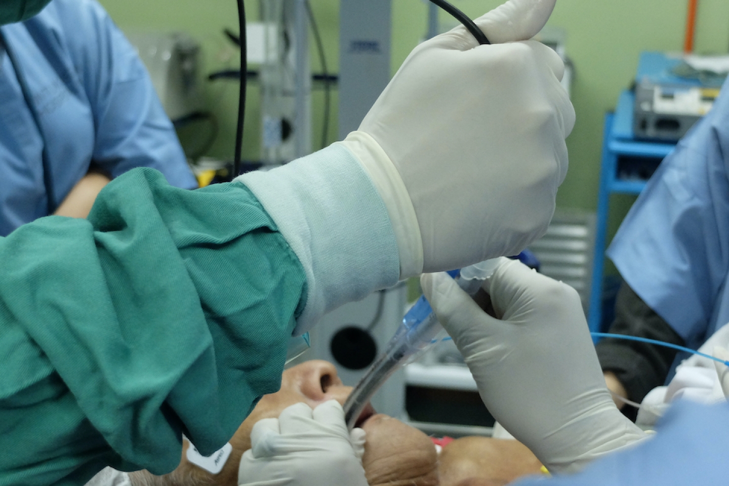
Pulmonologists have performed fiberoptic bronchoscopy to identify and biopsy lung lesions since 1967. One of the most commonly performed pulmonary procedures, bronchoscopy involves insertion of a thin camera through the mouth and into the windpipe to allow for a complete examination of the air passages while the patient is under anesthesia. This standard procedure has evolved from using tiny fiber optic cameras to digital cameras—but has faced limits.
“You can only go so far before the camera gets stuck or wedged and can’t advance further,” Dr. Shostak explains. “We now know that the area in the periphery of the lung is not good for traditional bronchoscopy. Many lesions require twists and turns to get to them, and our instruments couldn’t make those twists and turns because they were stiff, nor did we have any sort of image guidance to reach them.”
“The last decade has seen major advances in bronchoscopic technology”, Dr. Shostak adds. Pulmonologists are using ultrasound guidance when performing bronchoscopic biopsies of lymph nodes in the chest, and image-guided bronchoscopy, such as electromagnetic navigational bronchoscopy, to reach small nodules in the outer lung fields.
“Navigational bronchoscopy uses steerable, flexible instruments and GPS-like technology to allow us to reach nodules typically not accessible with traditional bronchoscopy,” Dr. Shostak explains. “This has significantly improved our ability to reach small nodules in the periphery of the lung.”
Despite these improvements, electromagnetic navigational bronchoscopy still has some drawbacks, including “CT-to-body divergence” – a discrepancy between the location of a nodule as seen on pre procedure scan and the actual location of the nodule during the procedure.
“In addition physicians lose image guidance at the time of bronchoscopic biopsy due to the design of the current navigational system,” Dr. Shostak adds.
For the last few months, Dr. Shostak and colleagues have used the ILLUMISITE™ platform to go beyond the traditional navigation technology. This upgraded system provides continuous GPS-style guidance; offers additional flexibility with a rotating fluoroscopy machine around the patient to avoid CT-to-body divergence; enhances hard-to-reach nodules; and contains extra sensors that provide continuous guidance during biopsy.
This system significantly increases ability to reach and biopsy small peripheral nodulesallowing pulmonologists to diagnose lung cancer earlier. Lymph node biopsies can be conducted in same setting to better understand whether patient’s cancer has spread.
Dr. Shostak adds, “We try to avoid any delays in care by offering a one-stop shop model. where patients can obtain both diagnosis and staging of their suspected lung cancer during one procedure.”
Subscribe to our mailing list to stay up to date on all the latest health news and important updates from Weill Cornell Medicine.